
Venogram
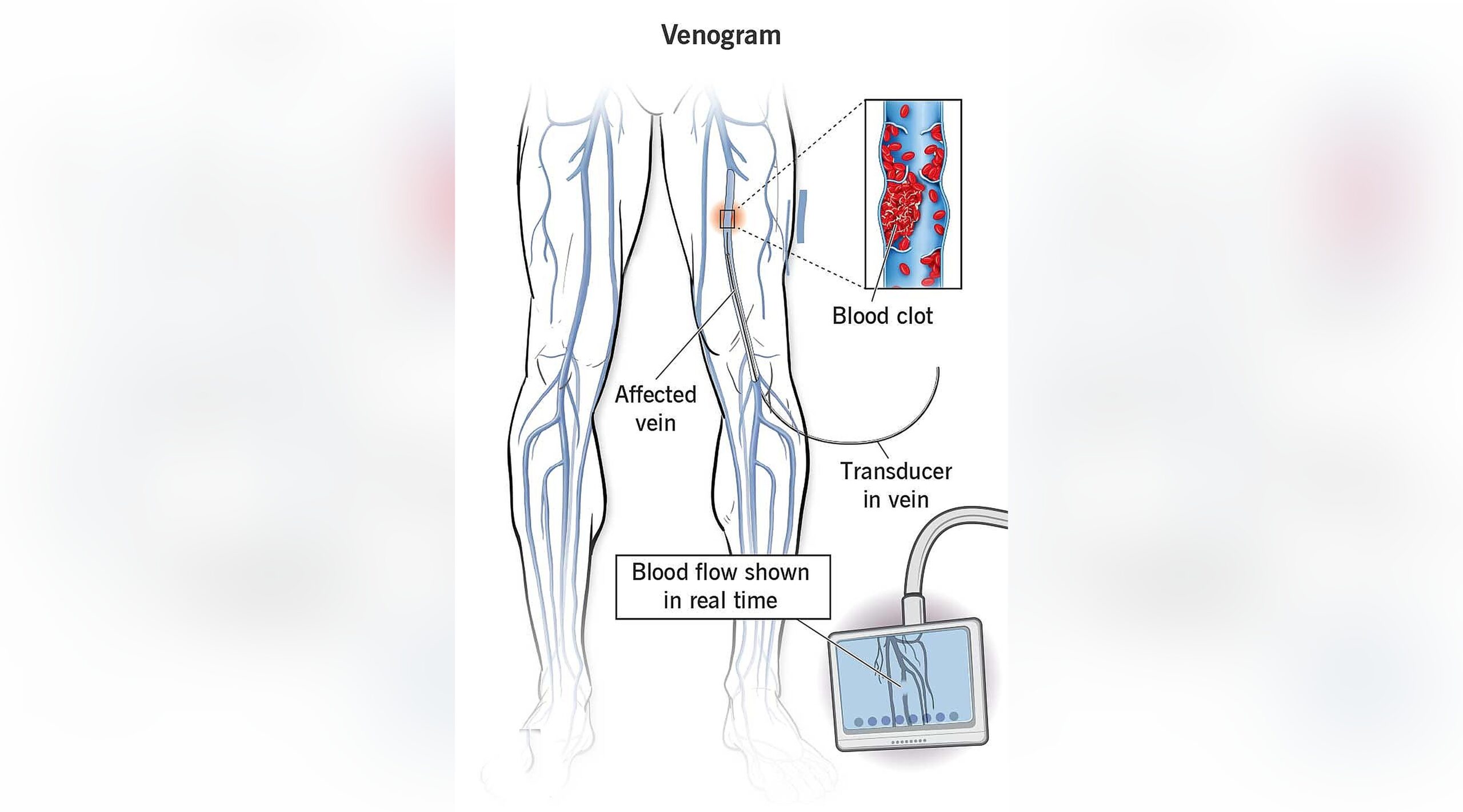
A Venogram is a minimally invasive imaging procedure used to evaluate the veins and blood flow within the venous system. By injecting contrast dye into a vein and capturing real-time X-ray images, physicians can identify blood clots, vein narrowing, blockages, abnormal blood flow patterns, and other venous disorders affecting the legs.
A venogram is often performed when detailed visualization of the veins is needed to diagnose complex venous conditions or to guide treatment planning for vascular interventions.
Why a Venogram Is Performed
Your healthcare provider may recommend a venogram if you have:
- Suspected deep vein thrombosis (DVT)
- Leg swelling of unknown cause
- Chronic venous insufficiency
- Leg pain related to venous disease
- Suspected vein narrowing or blockage
- Venous ulcers or non-healing wounds
- Abnormal findings on ultrasound studies
- Post-thrombotic syndrome
- Need for evaluation before venous intervention
- Recurrent symptoms despite previous treatment
The procedure provides detailed information about vein anatomy and blood flow that may not be visible with other imaging tests.
How the Procedure Works
Before the Procedure
Preparation may include:
- Review of your medical history and medications
- Blood tests to assess kidney function and clotting ability
- Discussion of allergies, particularly to contrast dye
- Temporary adjustment of blood-thinning medications if instructed
- Fasting for several hours before the procedure
- Placement of an intravenous (IV) line if needed
The procedure is usually performed in a hospital or outpatient vascular center.
During the Procedure
Line
- The patient lies comfortably on an X-ray table.
- The skin is cleaned and sterilized.
- A small needle or catheter is inserted into a vein, usually in the foot, ankle, leg, or groin.
- Contrast dye is injected into the vein.
- Real-time X-ray images are obtained as the dye travels through the venous system.
- Multiple images may be taken from different angles.
- The physician evaluates blood flow and vein anatomy.
- If necessary, additional imaging or venous pressure measurements may be performed.
- Once imaging is complete, the catheter is removed.
- Pressure is applied to the access site to prevent bleeding.
The procedure typically takes 30 to 60 minutes, depending on the complexity of the evaluation.
What to Expect During the Procedure
- You will remain awake throughout the procedure.
- Local anesthesia may be used at the access site.
- You may feel a brief warm sensation when the contrast dye is injected.
- Most patients experience minimal discomfort.
- Vital signs are monitored throughout the examination.
- Medical staff will provide instructions during image acquisition.
Benefits of a Venogram
- Provides detailed visualization of the veins
- Identifies blood clots and venous blockages
- Detects vein narrowing or compression
- Evaluates chronic venous insufficiency
- Assists with treatment planning
- Helps guide minimally invasive venous procedures
- Provides accurate assessment of complex venous conditions
- Can be combined with therapeutic interventions when needed
After the Procedure
Following the venogram:
- You may be observed for a short period before discharge.
- Mild soreness or bruising at the access site may occur.
- Drinking fluids is often encouraged to help eliminate contrast dye from the body.
- Most patients return to normal activities within 24 hours.
- Follow-up instructions will be provided based on the findings.
Your physician will review the results and discuss any recommended treatment options.
Risks and Safety
A venogram is generally safe, but potential risks include:
- Bruising or bleeding at the catheter site
- Infection
- Allergic reaction to contrast dye
- Blood clot formation
- Vein irritation or inflammation
- Kidney complications related to contrast dye
- Blood vessel injury
- Rare complications related to catheter placement
Your healthcare team takes extensive precautions to minimize these risks.
Understanding Your Results
A venogram may reveal:
- Normal venous blood flow
- Deep vein thrombosis (DVT)
- Venous narrowing or obstruction
- Chronic venous insufficiency
- Venous reflux
- Vein compression syndromes
- Post-thrombotic changes
- Abnormal venous anatomy
- Need for further treatment or intervention
The results help your physician determine the most appropriate treatment plan for your condition.
Frequently Asked Questions
Is a venogram painful?
Most patients experience only mild discomfort. Local anesthesia and modern catheter techniques help make the procedure comfortable.
How long does the procedure take?
Most venograms take approximately 30 to 60 minutes, although more complex evaluations may take longer.
Why would I need a venogram if I already had an ultrasound?
While ultrasound is often the first diagnostic test, a venogram provides more detailed visualization of certain veins and may be necessary when ultrasound findings are inconclusive or when intervention is being planned.
Can treatment be performed during the same procedure?
In some cases, if a significant blockage or narrowing is identified, additional venous interventions may be performed during the same session.
When can I return to normal activities?
Most patients resume normal daily activities within 24 hours, unless additional treatment is performed.
Schedule a Consultation
If you are experiencing leg swelling, venous pain, suspected blood clots, chronic venous insufficiency, or other symptoms of venous disease, a venogram can provide detailed information about your vein health and circulation. Contact our team today to schedule a consultation and learn whether this advanced diagnostic procedure is right for you.