
Venous Ultrasound Mapping
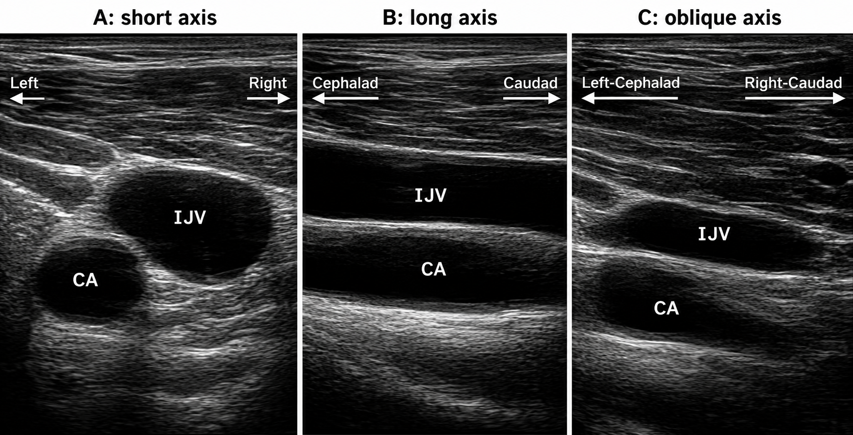
Venous Ultrasound Mapping is a noninvasive diagnostic imaging procedure used to evaluate the veins in the legs and create a detailed map of the venous system. Using high-frequency sound waves and Doppler technology, the examination helps identify abnormal blood flow, damaged vein valves, vein enlargement, blood clots, and other conditions that may contribute to venous disease.
This study is commonly performed before vein treatments such as endovenous ablation, Varithena®, sclerotherapy, or other venous interventions to help physicians develop an individualized treatment plan.
Why Venous Ultrasound Mapping Is Performed
Your healthcare provider may recommend venous ultrasound mapping if you have:
- Varicose veins
- Spider veins
- Chronic venous insufficiency
- Leg swelling
- Leg heaviness or fatigue
- Aching, throbbing, or cramping in the legs
- Skin discoloration related to venous disease
- Venous ulcers or slow-healing wounds
- Suspected blood clots
- Need for evaluation before vein treatment
The examination helps identify the source of venous reflux and provides valuable information for treatment planning.
How the Procedure Works
Before the Procedure
Preparation is usually simple and may include:
- Wearing loose, comfortable clothing
- Avoiding lotions or creams on the legs before the examination
- Bringing a list of medications and relevant medical history
- Following any instructions provided by your physician
Most patients do not require fasting or special preparation.
During the Procedure
Line
- The patient stands or lies on an examination table depending on the area being evaluated.
- A clear ultrasound gel is applied to the skin.
- A handheld ultrasound transducer is moved over the legs.
- Sound waves create real-time images of the veins.
- Doppler technology measures blood flow direction and velocity.
- The technologist evaluates vein size, valve function, and blood flow patterns.
- Areas of venous reflux or abnormal circulation are identified and documented.
- A detailed map of the venous system is created for physician review.
The examination typically takes 30 to 60 minutes.
What Venous Ultrasound Mapping Evaluates
The study may assess:
- Superficial veins
- Deep veins
- Perforator veins
- Venous valve function
- Blood flow direction
- Venous reflux (backward blood flow)
- Vein diameter and anatomy
- Blood clot formation
- Areas of venous obstruction
This information helps determine the most appropriate treatment approach.
What to Expect During the Examination
- The procedure is painless and noninvasive.
- No needles, incisions, or injections are required.
- You may be asked to change positions during the study.
- Gentle pressure from the ultrasound probe may be felt.
- You may hear blood flow sounds generated by Doppler technology.
- Most patients remain comfortable throughout the examination.
Benefits of Venous Ultrasound Mapping
- Noninvasive and painless
- No radiation exposure
- No contrast dye required
- Provides detailed evaluation of vein function
- Identifies the source of venous insufficiency
- Detects blood clots and venous abnormalities
- Assists with treatment planning
- Safe and accurate diagnostic tool
- Immediate return to normal activities
- Helps improve treatment outcomes
After the Examination
Following the ultrasound:
- The gel is removed from the skin.
- No recovery period is required.
- Patients can immediately resume normal activities.
- Images and findings are reviewed by a vascular specialist.
- Treatment recommendations are developed based on the results.
Your physician will discuss the findings and any recommended next steps.
Risks and Safety
Venous ultrasound mapping is considered extremely safe.
Potential considerations include:
- No exposure to radiation
- No surgical incisions
- No contrast material required
- No known harmful effects from diagnostic ultrasound
- No downtime or recovery period
The procedure is widely used because of its excellent safety profile and diagnostic accuracy.
Understanding Your Results
The examination may reveal:
- Normal venous blood flow
- Venous reflux (backward blood flow)
- Chronic venous insufficiency
- Enlarged or incompetent veins
- Varicose vein disease
- Blood clots within the veins
- Venous obstruction
- Areas requiring treatment
The results help your physician determine whether treatments such as endovenous ablation, Varithena®, sclerotherapy, or other vein procedures may be beneficial.
Frequently Asked Questions
Is venous ultrasound mapping painful?
No. The examination is painless and does not involve needles, injections, or surgery.
How long does the test take?
Most studies take approximately 30 to 60 minutes, depending on the complexity of the evaluation.
Does the test use radiation?
No. Ultrasound uses sound waves rather than radiation to create images.
Why is vein mapping important before treatment?
Vein mapping identifies abnormal veins and areas of venous reflux, allowing your physician to create a personalized treatment plan and improve treatment accuracy.
Can I return to work afterward?
Yes. Most patients return to work and normal daily activities immediately after the examination.
Schedule a Consultation
If you are experiencing varicose veins, leg swelling, aching, heaviness, skin changes, or other symptoms of venous disease, venous ultrasound mapping can provide valuable information about your vein health and circulation. Contact our team today to schedule a consultation and learn whether this noninvasive diagnostic evaluation is right for you.