
Vascular Ultrasound Evaluation
A Vascular Ultrasound Evaluation is a noninvasive imaging test used to assess blood flow through the arteries and veins of the legs. Using high-frequency sound waves, this examination provides real-time images of blood vessels and helps identify circulation problems such as narrowing, blockages, blood clots, or other vascular abnormalities.
Because it does not require needles, radiation, or contrast dye in most cases, vascular ultrasound is often one of the first tests used to diagnose Peripheral Artery Disease (PAD) and other circulatory conditions affecting the lower extremities.
Why a Vascular Ultrasound Evaluation Is Performed
Your healthcare provider may recommend a vascular ultrasound evaluation if you have:
- Leg pain while walking (claudication)
- Leg pain at rest
- Numbness or weakness in the legs
- Cold feet or lower extremities
- Non-healing wounds or ulcers
- Swelling in the legs
- Suspected Peripheral Artery Disease (PAD)
- Suspected blood clots
- Reduced or absent pulses in the legs
- Discoloration of the feet or toes
- A history of vascular disease
- Need for monitoring after a vascular procedure
The examination helps identify circulation problems and guides treatment planning.
How the Procedure Works
Before the Procedure
Preparation is usually simple and may include:
- Wearing comfortable, loose-fitting clothing
- Avoiding lotions or creams on the legs before the exam
- Following any specific instructions provided by your physician
- Bringing a list of medications and relevant medical history
Most vascular ultrasound examinations require little or no special preparation.
During the Procedure
- The patient lies comfortably on an examination table.
- A clear ultrasound gel is applied to the skin over the area being examined.
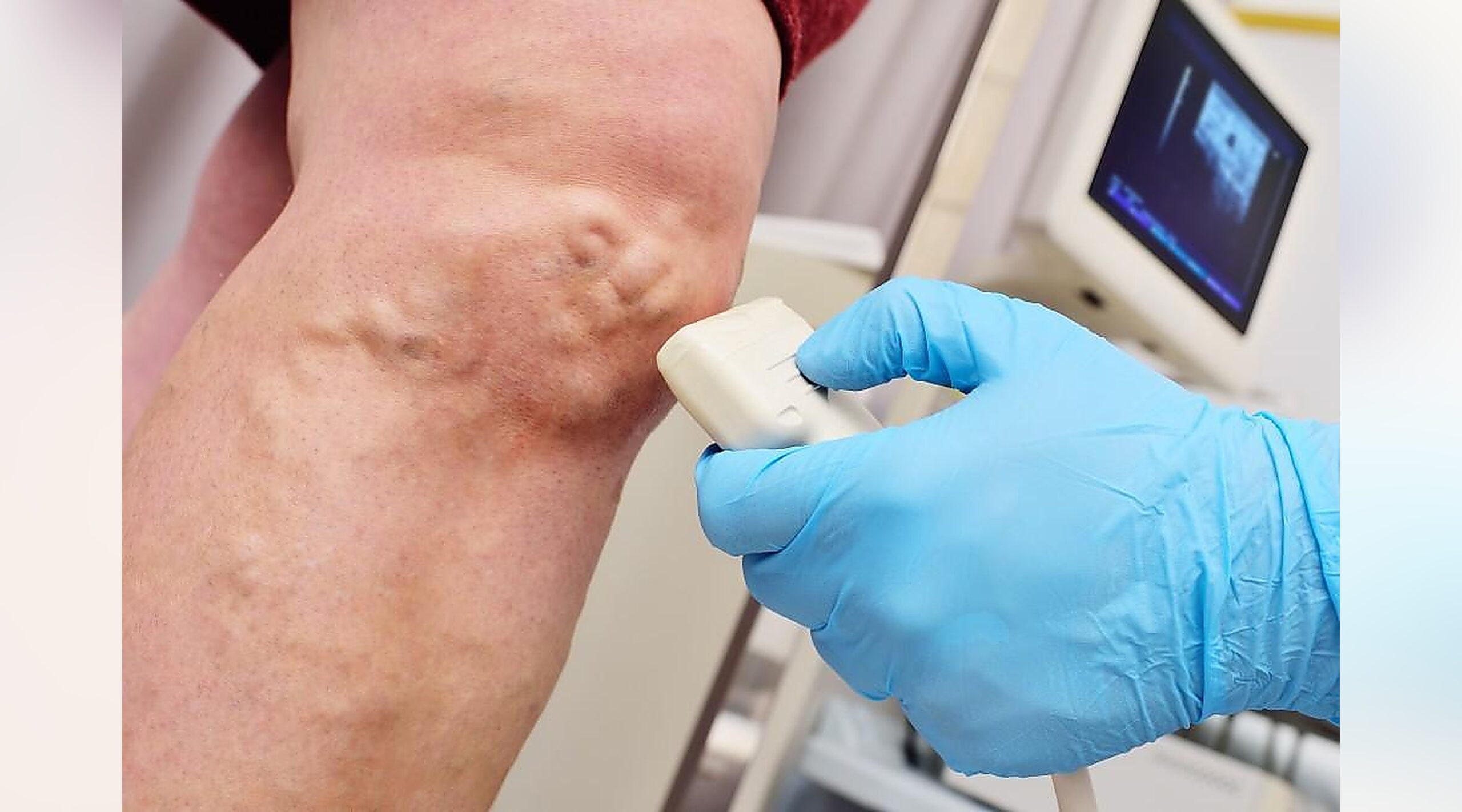
- A handheld device called a transducer is moved over the skin.
- The transducer sends sound waves into the body and receives returning echoes.
- Real-time images of the blood vessels appear on a monitor.
- Doppler ultrasound technology may be used to evaluate blood flow direction and speed.
- Multiple areas of the legs may be examined for a complete vascular assessment.
- The technologist records images and measurements for physician review.
The examination is painless and typically takes 30 to 60 minutes.
Types of Vascular Ultrasound Studies
Depending on your symptoms, your physician may recommend:
- Arterial Ultrasound – evaluates blood flow through the leg arteries
- Venous Ultrasound – evaluates veins and detects blood clots
- Doppler Ultrasound – measures blood flow velocity and direction
- Duplex Ultrasound – combines traditional ultrasound imaging with Doppler analysis
- Ankle-Brachial Index (ABI) Testing – may be performed alongside ultrasound to assess circulation
Your provider will determine which study is most appropriate for your condition.
What to Expect During the Examination
- The test is painless and noninvasive.
- No needles or incisions are required.
- You may be asked to change positions during the examination.
- Mild pressure from the transducer may be felt.
- You may hear blood flow sounds generated by Doppler technology.
- Most patients remain comfortable throughout the study.
Benefits of Vascular Ultrasound Evaluation
- Noninvasive and painless
- No radiation exposure
- No surgical incisions
- Provides real-time imaging of blood vessels
- Detects circulation problems early
- Helps diagnose Peripheral Artery Disease
- Identifies blood clots and vascular abnormalities
- Safe for most patients
- Quick outpatient procedure
- Useful for monitoring treatment outcomes
After the Examination
Following the ultrasound:
- The gel is removed from the skin.
- Patients can immediately resume normal activities.
- No recovery time is required.
- Results are reviewed by a physician specializing in vascular imaging.
- Additional testing or treatment may be recommended if abnormalities are detected.
Most patients can return to work, driving, and regular activities immediately.
Risks and Safety
Vascular ultrasound is considered extremely safe.
Potential considerations include:
- No exposure to ionizing radiation
- No known harmful effects from diagnostic ultrasound
- No contrast dye required in most examinations
- No recovery period or significant side effects
The procedure is widely used because of its excellent safety profile.
Understanding Your Results
The examination may reveal:
- Normal blood flow through the arteries and veins
- Narrowing or blockage of arteries
- Peripheral Artery Disease (PAD)
- Blood clots within veins
- Reduced circulation to the legs or feet
- Aneurysms or abnormal vessel enlargement
- Venous insufficiency or valve dysfunction
- Need for additional imaging or vascular intervention
Your physician will review the findings and discuss appropriate treatment recommendations.
Frequently Asked Questions
Is a vascular ultrasound painful?
No. The examination is painless and does not involve needles, injections, or surgical procedures.
How long does the test take?
Most vascular ultrasound evaluations take approximately 30 to 60 minutes.
Does the test use radiation?
No. Ultrasound uses sound waves rather than radiation to create images.
Can I return to normal activities afterward?
Yes. There are no activity restrictions, and most patients resume normal activities immediately.
When will I receive my results?
Results are typically reviewed by a physician and communicated within a few days, although timing may vary depending on your healthcare facility.
Schedule a Consultation
If you are experiencing leg pain, poor circulation, swelling, non-healing wounds, or other symptoms of vascular disease, a vascular ultrasound evaluation can provide valuable information about your blood flow and vascular health. Contact our team today to schedule a consultation and learn more about this safe, noninvasive diagnostic examination.